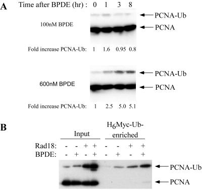
FIG. 1.
PCNA is monoubiquitinated after BPDE treatment. (A) Exponentially growing H1299 cells were treated with 100 nM (top) or 600 nM (bottom) BPDE. At different time points after BPDE treatment, nuclear fractions were prepared. After normalizing for protein content, nuclear samples were separated by SDS-PAGE, transferred to nitrocellulose, and analyzed by immunoblotting with anti-PCNA antibodies. (B) H1299 cells were infected with AdCon or AdRad18 for 24 h. The resulting cultures were transfected with H6M-Ub for 24 h and treated with BPDE (or left untreated). After 4 h, chromatin fractions from the cells were analyzed directly for PCNA levels. Alternatively, H6M-Ub-conjugated proteins were purified with cobalt-agarose resin prior to immunoblot analysis with anti-PCNA.

