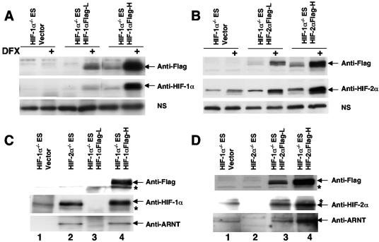
FIG. 6.
Endogenous HIF-2α forms a complex with ARNT in Hif-1α−/− ES cells. Establishment of Hif-1α−/− ES cells stably expressed low (L) or high (H) levels of Flag epitope-tagged HIF-1α protein (A) or HIF-2α protein (B) as assessed by Western blot analysis using anti-Flag or anti-HIF-α antibodies in the nuclear extracts derived from deferoxamine (DFX)-treated ES cells. Nonspecific protein bands (labeled NS) in the HIF-α Western blots served as loading controls. (C) Anti-HIF-1α antibody was used to immunoprecipitate HIF-1α protein in the nuclear extracts derived from hypoxia (1.5% O2)-treated ES cells. The amount of precipitated HIF-1α and coprecipitated ARNT protein was assessed using Western blot analysis. (D) Anti-HIF-2α antibody was used to immunoprecipitate HIF-2α in the nuclear extracts derived from hypoxia (1.5% O2)-treated ES cells. ARNT protein is coprecipitated with the HIF-2α protein in Hif-1α−/− ES cells (lane 1). Nonspecific protein bands (labeled by an asterisk) in anti-Flag Western blots (for panels C and D) exhibited similar intensities in all lanes.

