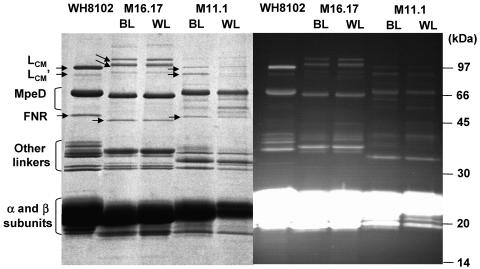
FIG. 7.
Coomassie blue-stained (left) and UV-visualized (right) LiDS-PAGE gels showing the linker composition of intact PBSs from Synechococcus species strains M11.1 and M16.17 grown in blue (BL) and white light (WL). The corresponding profile obtained for the chromatically nonadapting Synechococcus sp. strain WH 8102, in which all the linkers have been firmly identified (33), are shown for comparison. Note that the latter profile differs from the previously published one by an artifactual doubling of the MpeE band at ca. 38 kDa. Double arrows indicate the positions of LCM and LCM′ linker polypeptides (both of which fluoresce blue under UV light), and the single arrow indicates the likely position of FNR in each strain. Minor bands located between the FNR and MpeD in M11.1 are likely proteolytic fragments of MpeD.

