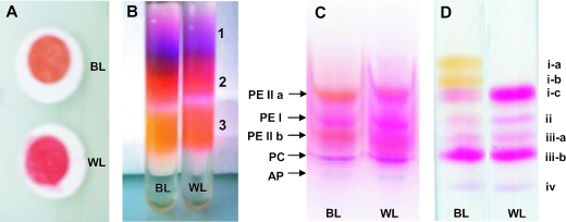
FIG. 8.
Different purification steps of phycobilisome components from Synechococcus sp. strain M16.17 cells grown in blue light (BL) and white light (WL). Filtered whole cells (A); dissociation of soluble proteins on continuous sucrose gradient showing the different fractions 1, 2, and 3 (B); native IEF gel of soluble proteins showing separated PBP complexes, including two PEII fractions (a and b) (C); and denaturing IEF showing separated PBP polypeptides (D). See the text for details.

