
FIG. 5.
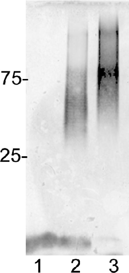
Immunoblot of C. jejuni waaC capsular material from a 12% SDS-PAGE gel. Lane 1, 81-176 kpsM mutant (2); lane 2, 81-176; and lane 3, 81-176 waaC mutant. The positions of protein markers in kDa are shown at the left. The LOS cores of the 81-176 kpsM mutant and the wild type are visible at the bottom of the immunoblot; the core of the waaC mutant is not visible.