FIG. 1.
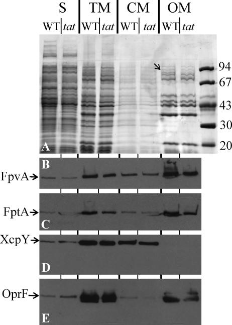
FpvA is transported into the outer membrane in a Tat-independent manner. Bacterial cell culture of wild-type PAO1 (WT) and its isogenic tatABC mutant (tat) grown in iron-limited medium were subjected to subcellular fractionation. S, soluble fraction containing cytoplasmic and periplasmic protein; TM, whole cell envelope; CM, cytoplasmic membrane; OM, outer membrane. The equivalent of 0.1 OD600 unit of the cultures was loaded onto a 9% SDS-PAGE gel and then stained with Coomassie blue (A). Alternatively, proteins were blotted onto nitrocellulose and revealed using anti-FpvA antibody (B), anti-FptA antibody (C), anti-XcpY antibody (D), or anti-OprF antibody (E). Molecular mass markers (kDa) are shown on the right. The position of FpvA, identified by mass spectrometry, is shown by an arrow in panel A.

