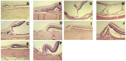
FIGURE 3.
Histologic examination of the margins of colobomas in pediatric patients. In general, the inner retinal layer extends centrally, and the outer layer reverts to meet the laterally displaced retinal pigment epithelium (RPE). The perverse orientation of the photoreceptors is shown (A, B, C, E), as are the radial glia oriented parallel to scleral fibers forming a triangle between the inner and outer retina (D, E, F, I). There is exceptional vascular support of the margin (D), vitreous traction (G), and schisis of the transition to intercalary membrane (H). There is choroidal thickening and RPE hyperplasia (I, J), as well as subchoroidal invasion by the retina (I), which resembles a pocket. Rosettes are part of the margin (A, C). A, Patient 6, hematoxylin-eosin, ×10. B, Patient 21, hematoxylin-eosin, ×10,. C, Patient 18, hematoxylin-eosin, ×10. D, Patient 20, hematoxylin-eosin, ×10. E, Patient 18, hematoxylin-eosin, ×4. F, Patient 23, hematoxylin-eosin, ×10. G, Patient 2, hematoxylin-eosin, ×4. H, Patient 1, hematoxylin-eosin, ×4. I, patient 11, hematoxylin-eosin, ×10. J, Patient 20, hematoxylin-eosin, ×10.

