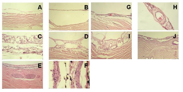
FIGURE 4.
Histologic examination of the center of colobomas in adult patients. In general, the variably atrophic intercalary membrane is directly apposed to the sclera. Neuronal atrophy (A through J), schisis (D, I), and arteriosclerosis of intercalary vessels (G through J) are consistent with aging. The thinned sclera contains nerves (E, F). A, Patient 7, hematoxylin-eosin, ×10. B, Patient 8, hematoxylin-eosin, ×10. C, Patient 8, hematoxylin-eosin, ×40. D, Patient 14, hematoxylin-eosin, ×10. E, Patient 26, hematoxylin-eosin, ×10. F, Patient 14, hematoxylin-eosin, ×40. G, Patient 8, hematoxylin-eosin, ×10. H, Patient 8, hematoxylin-eosin, ×40. I, Patient 14, hematoxylin-eosin, ×10. J, Patient 22, periodic acid–Schiff stain, ×10.

