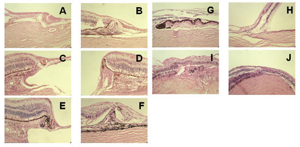
FIGURE 5.
Histologic examination of the margin in adult patients. In general, there is an abrupt termination of the retinal pigment epithelium and choroid, which are often hyperplastic and thickened. Both are centrally displaced, eliminating the point of reversal and locus minoris resistentiae and thus adding to structural stability. Moreover, abundant vascular supply of the margin is seen (A, C, D, E). Choroidal thickening and pockets anchor the intercalary membrane and form a seal (B through F), and intercalary schisis and hole formation reduce traction on the margin (C, E). Drusen-like deposits are consistent with aging (G). In smaller colobomas, well-vascularized intercalary membranes show preservation of inner layers (I, J). A, Patient 7, hematoxylin-eosin, ×10. B, Patient 8, hematoxylin-eosin, ×4. C, Patient 8, hematoxylin-eosin, ×10. D, Patient 8, hematoxylin-eosin, ×10. E, Patient 8, hematoxylin-eosin, ×10. F, Patient 14, hematoxylin-eosin, ×4. G, Patient 22, hematoxylin-eosin, ×10. H, Patient 15, hematoxylin-eosin, ×10. I, Patient 9, hematoxylin-eosin, ×4. J, Patient 26, hematoxylin-eosin, ×10.

