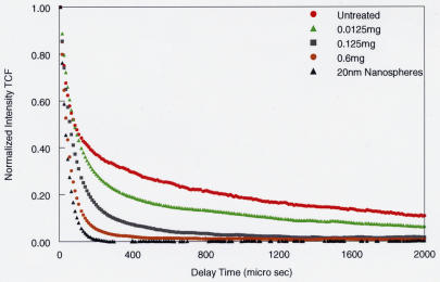
FIGURE 8.
Dynamic light-scattering (DLS) measurements in porcine vitreous 90 minutes after microplasmin injection 4 mm posterior to the lens. The effects of microplasmin on porcine vitreous are compared to untreated vitreous and a solution of 20-nm polystyrene beads. DLS measurements were performed at a site 4 mm posterior to the anterior vitreous cortex, simulating the intended site of intravitreal injections in vivo. The time correlation function (TCF) curve (see Appendix A) for untreated whole vitreous (red circles) demonstrates the two components previously identified as arising from the major macromolecules hyaluronan and collagen (Figure 6). With increasing doses of microplasmin, there is a decrease in the slope of the TCF, signifying breakdown of the larger vitreous macromolecules, most likely collagen, followed by disappearance of the smaller, more flexible macromolecules, most likely hyaluronan, ultimately approaching the TCF of a pure solution of nanospheres (black triangles.) The smaller particles are likely the breakdown products of the larger vitreous macromolecules induced by microplasmin.

