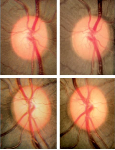
FIGURE 3.
Photomicrographs of the endothelin-1 treated (top panels) and contralateral control (bottom panels) optic nerves in one monkey (monkey 8) taken at baseline and at the end of 6 months (final, top right and bottom right panels) of ischemia. This monkey has 16% axonal loss of the entire optic nerve, with the most severe loss in the nasal and superior nasal regions. There is no evidence of diffuse or focal pallor or optic disc edema, findings that are commonly associated with anterior ischemic optic neuropathy. Reprinted with permission from Archives of Ophthalmology.132

