
Figure 5.—
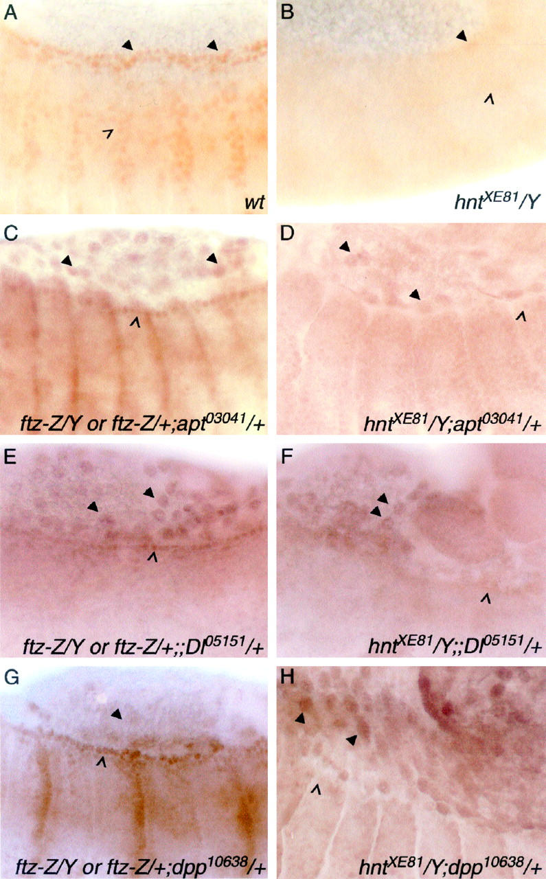
HNT regulates candidate interacting genes tissue autonomously and nonautonomously in the embryo. (A and B) Wild-type (A) and hntXE81 (B) stage 14 embryos showing APT protein in the dorsal vessel (arrowheads) and the tracheal system (open arrowhead). APT expression can be seen to be very reduced in the hnt mutant. (C–H) Expression reported by lacZ enhancer trap lines detected with anti-β-galactosidase antibody. The left column shows embryos with one copy each of the lacZ insertion and the FM7, ftz-lacZ balancer chromosome. The latter distinguishes these embryos from their hntXE81 male siblings (right column). apontic (apt) expression in the leading edge (open arrowhead in C) is almost absent in hnt mutant embryos (open arrowhead in D), whereas the amnioserosal expression is not altered (arrowheads in C and D). A similar result is seen with Delta (Dl) expression (E vs. F; leading edge, open arrowheads; amnioserosa, arrowheads). dpp expression is upregulated in the amnioserosa of hnt mutant embryos (arrowheads in G vs. H) while epidermal leading edge expression is nonautonomously reduced (open arrowheads in G and H).