
Figure 4.
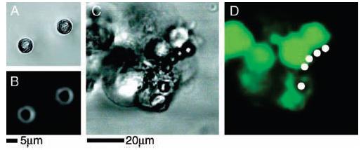
Adherence of HEK293 cells to polystyrene beads covered with chemically coupled neuroligin-1. White circles denote the positions of polystyrene beads in fluorescence images. (A, B) HEK293 cells not transfected with β-neurexin; no beads are observed to adhere to the cells and have fallen away from the coverslip after inversion of the chamber. (C) Bright field image of polystyrene beads, coupled to neuroligin-1, adhering to HEK293 cells expressing β-neurexin. In all cases, both transfected and nontransfected cells remained adhered to the glass coverslip. (D) Fluorescence image of polystyrene beads, coupled to neuroligin-1, and adhering to β-neurexin-expressing HEK293 cells.