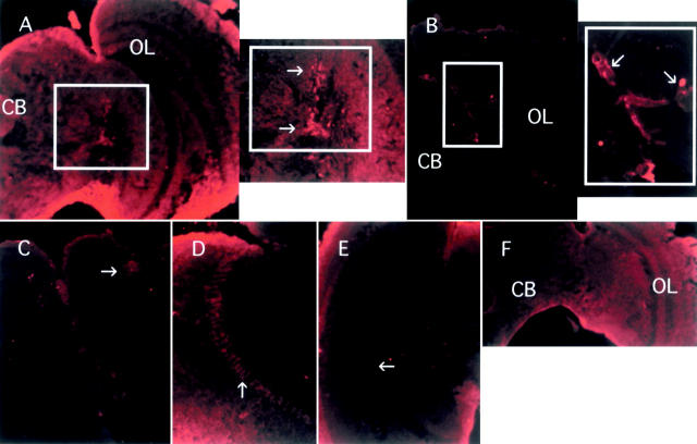
Figure 4.—
Immunoreactivity to anti-PER antibody detected in frontal sections of male B. tryoni brain. Similar results were seen in female brain. Arrows indicate the positive signals. Staining at the edge of the section is due to background. (A) Central brain and optic lobe with framed lateral region. The enlarged lateral region is shown on the right. CB, central brain; OL, optic lobe. The fly was sampled in the afternoon at 16:00. (B) Central brain and optic lobe from a fly sampled at night (23:00) with enlarged lateral region on the right showing more intense PER staining compared to A. (C) Dorsal region. The fly was sampled in the night at 23:00. (D) Optic lobe showing staining of glial cells. The fly was sampled at 23:00. (E) Optic lobe of a fly sampled at 16:00. Note extremely weak staining in glial cells (arrow). (F) Negative control. Staining of a fly brain sampled at 23:00 without anti-PER antibody. Diffuse background fluorescence is present, but there is no evidence of cell-specific staining.

