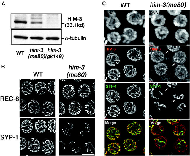
Figure 1.—
Cohesin-containing chromosome axes with reduced levels of HIM-3 protein support limited synapsis in the him-3(me80) mutant. (A) Western blots containing lysates from equal numbers of adult wild-type (WT) worms, him-3(me80) worms, and worms carrying the deletion allele him-3(gk149) (Couteau et al. 2004), probed with anti-HIM-3 and anti-α-tubulin antibodies. As the him-3(me80) mutation resides outside the fragment used to produce the anti-HIM-3 antibody, the reduction in the 33-kD HIM-3 band in the him-3(me80) lane reflects reduced levels of HIM-3 protein rather than absence of epitopes. (B and C) Immunostaining of REC-8 and SYP-1 (B) and HIM-3 and SYP-1 (C) in nuclei from the mid-pachytene regions of whole-mount WT and him-3(me80) germ lines. Images are projections of 3-D data stacks encompassing whole nuclei. In B, him-3(me80) shows more numerous stretches of REC-8 and fewer stretches of SYP-1 than seen in WT. In C, HIM-3 and SYP-1 immunostaining are shown in conjunction with DAPI staining of chromatin. For the him-3(me80) panels, the detection threshold for the anti-HIM-3 signal was lowered to permit imaging of the greatly reduced HIM-3 signal intensity in this mutant; consequently the background appears higher than in WT in this channel. The reduced HIM-3 signal is broadly distributed along all chromosomes in the him-3(me80) mutant; SYP-1 stretches coincide with a subset of HIM-3 stretches, typically including those with brighter HIM-3 signals (arrows). In the merged image, HIM-3 is shown in red and SYP-1 is shown in green. Bars, 5 μm.

