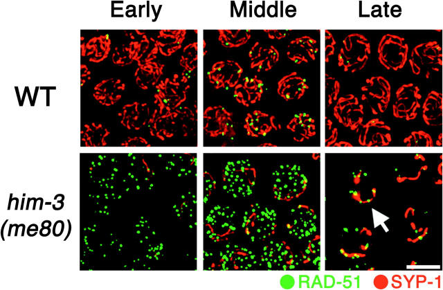
Figure 6.—
Simultaneous visualization of RAD-51 foci and SC central region protein SYP-1. Immunostaining of RAD-51 (green) and SYP-1 (red) in pachytene region nuclei from whole-mount germ lines of wild-type and him-3(me80) hermaphrodites is shown. Images are projections of 3-D data stacks encompassing whole nuclei; for each genotype, the three consecutive panels show different regions of the same germ line. Left, the early pachytene region, where very few SYP-1 stretches are seen in the him-3(me80) mutant and RAD-51 foci are already more abundant than in wild type. Middle, the mid-pachytene region, where RAD-51 foci in the him-3(me80) mutant appear larger than those in the left panel; some RAD-51 foci show colocalization with SYP-1 stretches, but foci are abundant on regions that lack SYP-1. Right, late pachytene region, where RAD-51 foci are greatly diminished in number compared to earlier stages but are preferentially retained at SYP-1 stretches (arrows) in the him-3(me80) mutant. Bar, 5 μm.

