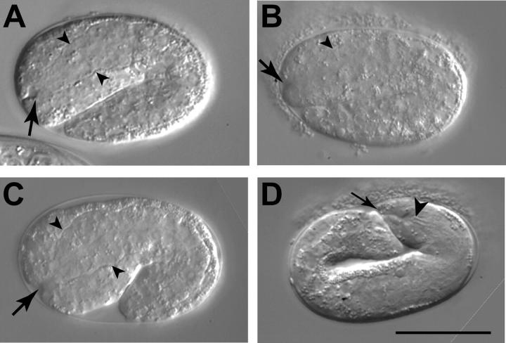
Figure 4.—
Abnormal morphology at the junction of the hypodermis and pharyngeal primordium in embryos. (A) A pronounced invagination (arrow) at the anterior end of a mec-8; sym-3 mutant embryo. The basement membrane of the pharyngeal primordium is evident (arrowheads). (B) An apparently normal invagination (arrow) at the anterior end of a mec-8(−); sym-3(+) sibling of the embryo in A. The focal plane with the maximum invagination is shown. The dorsal part of the pharyngeal primordium can be seen (arrowhead). (C) The invagination of a wild-type N2 embryo. (D) A mec-8; sym-3 embryo later in development exhibiting extreme abnormality at the anterior end. The buccal cavity (arrowhead) is grossly recessed from the anterior tip of the embryo (arrow). A canal may exist between the anterior tip and the buccal cavity. In contrast, the anterior ends of normal embryos at this stage resemble those of the larvae shown in Figure 3, D and F. Bar, 25 μm.

