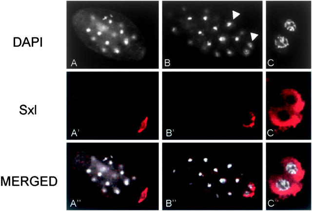
Figure 8.—
Distribution of Sxl protein in preblastoderm stage embryos of S. coprophila. (A and B) DAPI staining and indirect immunolabeling with anti-Sxl antibody (in red) of a whole embryo showing the germ nuclei (arrow) and the somatic nuclei (arrowhead). (C) Partial view of germ nuclei surrounded by Sxl protein.

