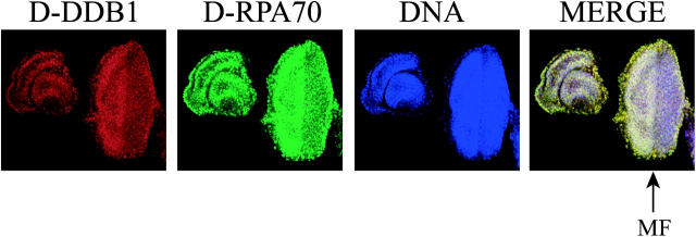
Figure 4.—
Distribution of D-DDB1 in the eye imaginal disc. The anterior of the discs is on the left and the posterior is on the right. Triple labeling (D-DDB1 in red, DNA in blue, and D-RPA70 in green) shows, in merged views: red, D-DDB1; blue, DNA; green, D-RPA70; and yellow, D-DDB1 and D-RPA70. Arrows indicate the position of the morphogenetic furrow (MF). D-DDB1 is especially abundant anterior to the MF. Essentially, similar patterns were found for both D-DDB1 and D-RPA70.

