
Figure 3.—
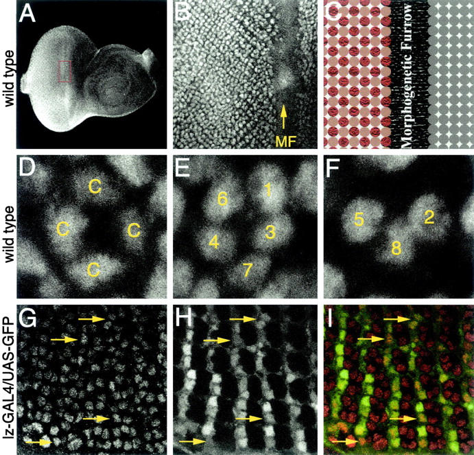
CBP is expressed in all cells of the developing eye imaginal disc. Confocal images of third instar imaginal discs are shown. Genotypes are listed at the left of each row. (A and B) Low and high magnification view of CBP expression ahead of and behind the morphogenetic furrow (MF). (C) Schematic of cells within the eye disc. Gray circles represent cells ahead of the furrow. Red circles represent ommatidial clusters. Brown circles represent intervening cells. (D–F) CBP is present in cone (c) and photoreceptor cells (1–8). (G–I) CBP is present in the intervening cells. (G) CBP; (H) lz-GAL4/UAS-GFP; and (I) merge of G and H. Yellow arrows indicate intervening cells. Anterior is to the right.