
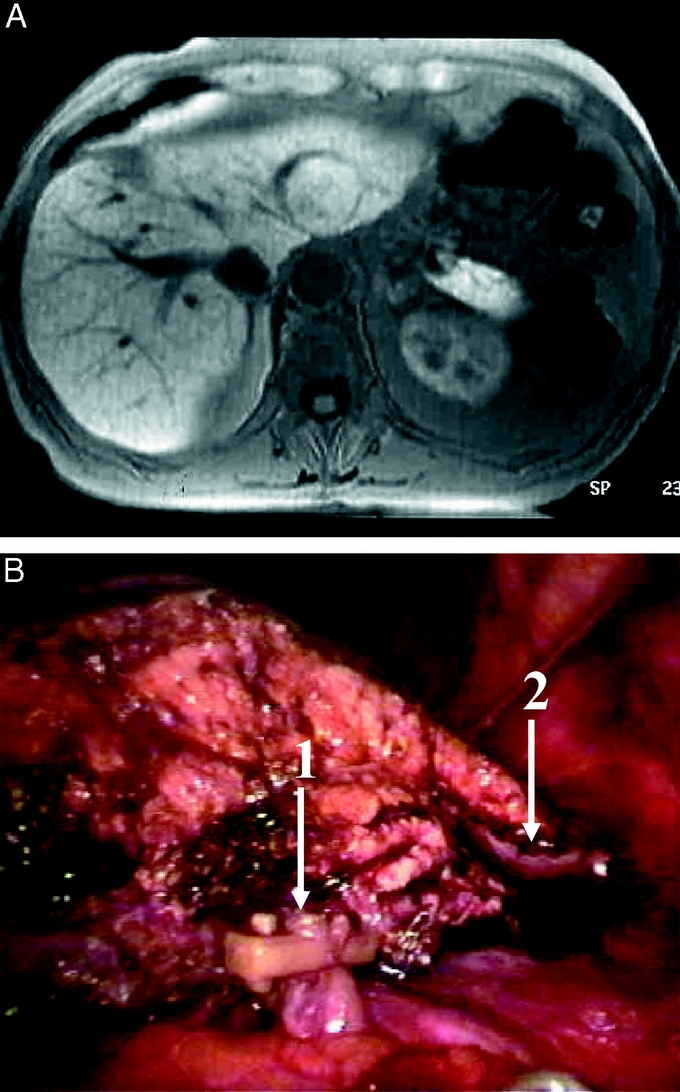
FIGURE 2. Left lateral sectionectomy for a 3-cm HCC in segment 2. Preoperative magnetic resonance (above) and operative view of completed resection (below). Arrow 1 shows a locked clip on a left arterial branch, and arrow 2 shows the stapled stump of the left hepatic vein.