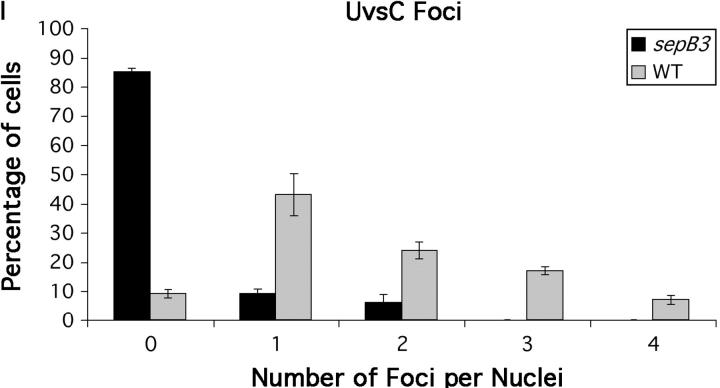
Figure 4.—
UvsC localizes to DNA-damage-induced subnuclear foci. (A, B, E, and F) Wild-type (ASG17) hyphae. (C, D, G, and H) sepB3 (ASG19) hyphae. Strains were grown in YGV for 14 hr at 28° and then left untreated (A–D) or exposed to 10 μg/ml PLM (E–H) and examined by immunofluorescence microscopy. UvsC-FLAG was detected using anti-FLAG antibodies (A, C, E, and G), and nuclei were stained using Hoechst 33258 (B, D, F, and H). (I) The number of UvsC-FLAG foci per nucleus was determined for wild-type (ASG17) and sepB3 (ASG19) hyphae treated with 10 μg/ml PLM. UvsC-FLAG was detected as described above, and hyphae were examined by immunofluorescence microscopy. For each sample, 100 nuclei were examined. Bars, 4 μm.

