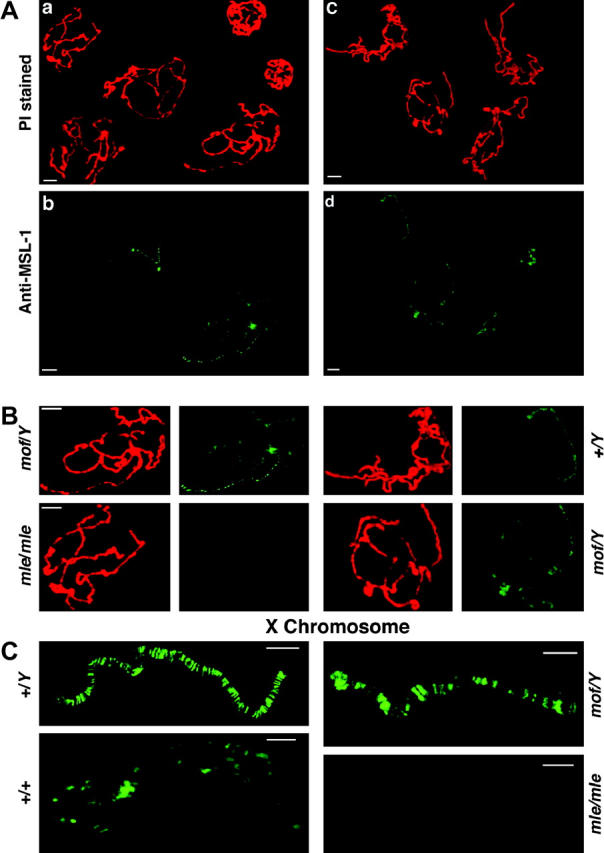
Figure 5.—

Distribution of the MSL complex in the mof mutant as revealed by MSL-1 labeling. (A) (a) A mixture of polytene chromosomes from mof males and mle/mle females stained with propidium iodide (PI). (b) The same nuclei probed for MSL-1. The females have no detectable MSL labeling on their chromosomes (Bhadra et al. 1999) and serve as a background control. The mof males show a destabilization of the MSL complex such that a low level of binding is now present on the autosomes, although a stronger labeling still occurs on the X. (c) A mixture of polytene chromosomes from mof and normal males stained with PI. (d) The same nuclei probed for MSL-1. The normal males show the usual strong labeling on the X chromosome and no detectable autosomal labeling. In contrast, the mof males exhibit some degree of MSL labeling on the autosomes. (B) Magnified views for comparison of single nuclei from the two mixtures in A. (C) Magnified views of the X chromosome from four genotypes. The normal male (+/Y) has strong labeling on the X. The normal female (+/+) (from a labeling not shown above) has a lower level of labeling, which is similar to the autosomes in the same nucleus (Hiebert and Birchler 1994). The mof male (mof/Y) retains strong binding at the high-affinity sites and weaker binding between them. The mle/mle female (mle/mle) shows no detectable labeling and illustrates that the signal between the strongly bound sites in the mof male is above background.
