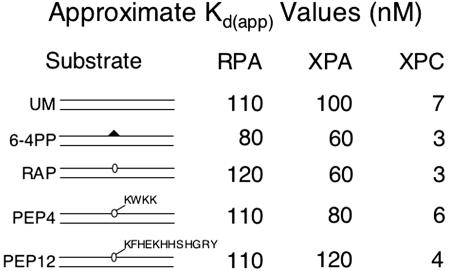
Fig. 4.
DNA binding by RPA, XPA, and XPC. Substrate DNA (0.05 nM) was incubated with increasing amounts of RPA (50–200 nM), XPA (25–200 nM), or XPC (2–10 nM) in the presence of 0.1 nM undamaged DNA. Binding was analyzed by electrophoretic mobility-shift assay and quantified by PhosphorImager analysis. Approximate Kd(app) values were estimated by visual examination of binding isotherms to determine the concentration (nM) at which 50% substrate was bound. UM, unmodified; 6–4PP, T[6-4]T photoproduct; PEP4, DNA–KWKK crosslink; PEP12, DNA–KFHEKHHSHGRY crosslink.

