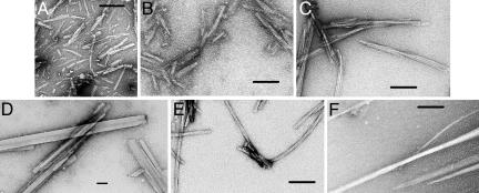
Fig. 1.
Electron micrographs showing fibrils (A–C and E) and needle-shaped microcrystals (D and F) formed by insulin and β2m and hexameric peptides from their sequences. (A) β2m91:KIVKWD. (B) β2m83:NHVTLS. (C) Full-length β2m. (D) β2m62:FYLLYY. (E) Full-length insulin. (F) IB12:VEALYL. The number following the protein name is the position in the protein sequence of the first residue. The sequence of the hexameric peptide follows. The length of the calibration bars is 100 nm.

