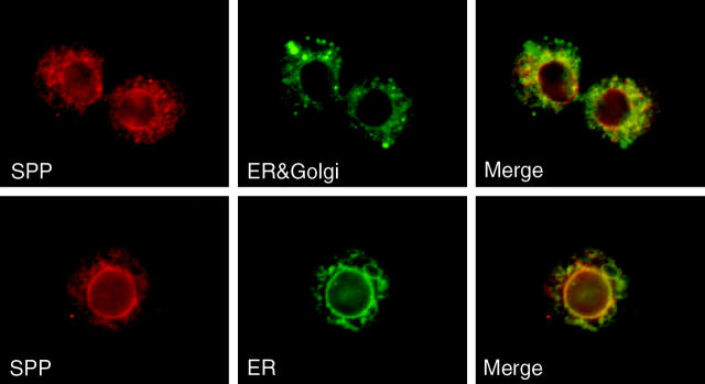
Figure 2.—
Immunofluorescence of Schneider-2 cells expressing MYC-tagged Drosophila SPP. Drosophila SPP that was tagged on either the N terminus (top) or the C terminus (bottom) revealed perinuclear and reticular localization. Left, SPP (red); middle, KDEL-receptor-GFP fusion protein (top) and calreticulin-GFP-KDEL fusion protein (bottom), labeled ER + Golgi and ER, respectively (green); right, merged images.

