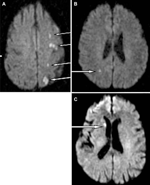
FIGURE 2.

DWI scans from three patients (A–C) with significant DWI lesions. Only one patient (A) had cognitive dysfunction; the others (B and C) did not. Of note, even though scans of the brain from the vertex through the brain stem including the cerebellum were analyzed, the images shown are for those parts of the brain that show the most prominent lesions. Lesions indicated by white arrows.
