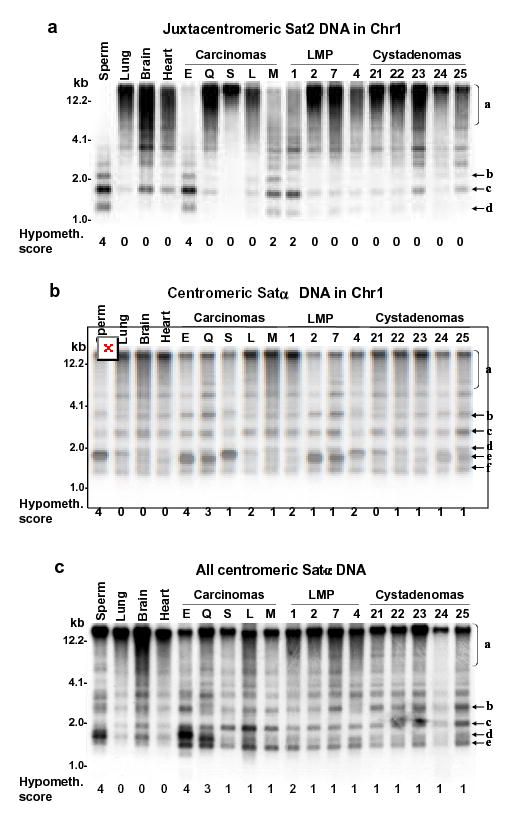
Fig. 1. Southern blot analysis of satellite DNA hypomethylation in ovarian epithelial tumors.

Examples of satellite DNA hypomethylation in BstBI digests of ovarian carcinomas in a single blot hybridized three times. Normal postnatal somatic DNAs and sperm DNA are the hypermethylated and hypomethylated standards, respectively. (A) juxtacentromeric Chr1 Sat2 probe and (B) centromeric Chr1 Satα probe under high-stringency hybridization conditions; (C), centromeric Chr1 Satα probe under low-stringency conditions that allow hybridization to DNA from most of the centromeres. Scoring of hypomethylation was by phosphorimager quantitation of <4-kb vs. >4-kb signal and visual comparison of bands b, c, d, e, and f to region a.
