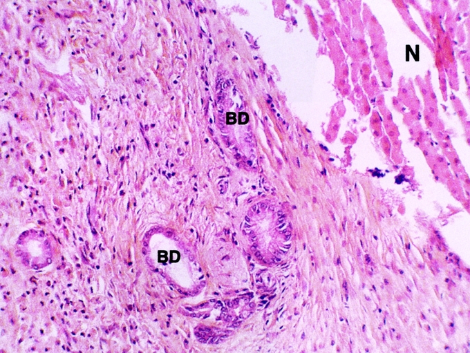
FIGURE 3. Histologic appearance of bile duct (BD) after hepatic radiofrequency (RF) necrosis with hepatic pedicle clamping. The bile ducts were cooled with cold 5% glucose isotonic serum. Hepatic removal was performed 3 weeks after RF (hematoxylin and eosin stain). The bile ducts in contact with the necrotic area were preserved. Necrosis (N) of hepatic parenchyma (original magnification ×20).
