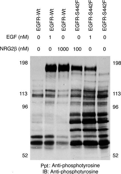
Figure 8. EGFR-Wt and the EGFR-S442 mutant couple with distinct signalling pathways.
32D cells that express the EGFR-Wt or the EGFR-S442F mutant were treated with PBS, 1 nM EGF or 1000 nM NRG2β. Protein tyrosine phosphorylation was analysed by immunoprecipitation and immunoblotting with an anti-phosphotyrosine antibody (4G10). The position of the molecular mass standards is indicated. The band of approx. 180 kDa is presumed to be tyrosine-phosphorylated EGFR.

