
FIGURE 6:
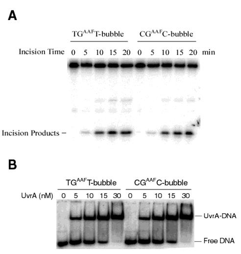
Incision and binding of AAF-DNA bubble substrates. (A) The AAF-DNA bubble substrates (B3′, Figure 1B) with the adduct sequences TG*T and CG*C were incised by UvrABC nuclease. The 5′-terminally labeled substrates were incubated with UvrABC (UvrA, 15 nM; UvrB, 250 nM; and UvrC, 100 nM) in the UvrABC buffer at 37 °C for various periods. The incision products were analyzed on a 12% polyacrylamide sequencing gel. (B) Binding of UvrA to the same bubble substrates. The 5′-terminally labeled DNA substrates were incubated with varying concentrations of UvrA at 37 °C for 15 min in the UvrABC buffer without ATP. The binding products were analyzed on a 3.5% native polyacrylamide gel.