
FIGURE 1.
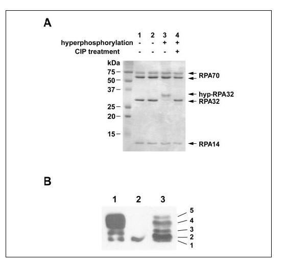
In vitro phosphorylation of RPA by DNA-PK. A, the phosphorylation reaction was conducted as described under “Experimental Procedures.” Proteins including untreated RPA (lane 1), mock-treated RPA (lane 2), hyp-RPA (lane 3), and calf intestinal phosphatase (CIP)-treated hyp-RPA (lane 4) were visualized by 14% SDS-PAGE and Coomassie Blue staining. B, immunoblot of RPA hyperphosphorylated by DNA-PK (lane 1), RPA (lane 2), and RPA in UV-irradiated HeLa cells (lane 3). To prepare the RPA in UV-irradiated HeLa cells, whole cell lysates were prepared from HeLa cells at 8 h after irradiation with 20 J/m2 UV. Proteins were separated on a 10% SDS-PAGE followed by Western blotting using an antibody specific for RPA32.