
Figure 4:
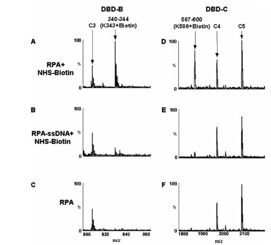
MALDI-TOF analysis of protection of lysine residues. The peptide peak containing modified K343 of DBD-B (A) is absent in the presence of the dT30mer (B). Similarly, K588 is located in DBD-C and is biotinylated in the absence of ssDNA (D) but is protected from modification by direct contact with the dT30mer (E). Panels C and F show the spectra of RPA without treatment of biotin. The unmodified peaks C3, C4, and C5 all serve as controls.