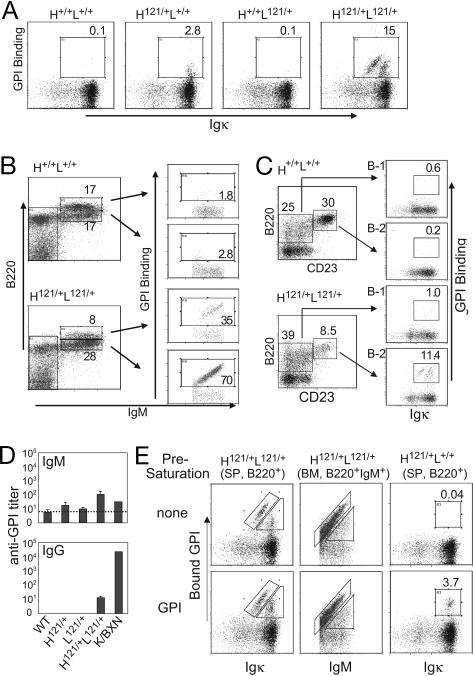
Fig. 1.
Anti-GPI B cells in 121 knock-in mice. (A) B220+ splenocytes from adult knock-in mice (H121/+L121/+) and control littermates stained with anti-Igκ and GPI-biotin. (B) Bone marrow B cells of adult knock-in (H121/+L121/+) and wild-type (H+/+L+/+) mice, stained with anti-B220, anti-IgM, and GPI-biotin, and gated on lymphocyte population. (C) Igκ and GPI profiles for B-1 (B220loCD23−) and B-2 (B220hiCD23+) lineages of the peritoneal cavity lavage cells. (D) ELISA titers of anti-GPI IgM or IgG antibodies in the serum of H121/+L121/+ and control littermates (n = 3–5). Titer is defined here as the serum dilution that gave an optical density of 2× background. A pooled serum from K/BxN arthritic mice was included as a reference. (E) The extent of occupancy by self-antigen of anti-GPI surface receptors was analyzed by staining cells directly (Upper), or after preincubation with saturating amounts of GPI (100 μg/ml; Lower). Cytometry profiles representative of 3–10 independent mice.

