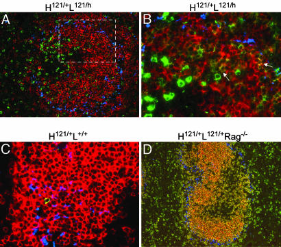
Fig. 5.
Localization of GPI-binding B cells in spleen. Spleen sections from various mice were stained with FITC-GPI (shown in green) and Moma-1 antibody (shown in blue to mark the boundary of the marginal zone). (A) Spleen sections costained with hCκ (shown in red) (×16 objective). (B) Magnification of the area marked by the white rectangle in A (×40 objective). GPI-binding cells appear in green, and allelicly included cells coexpressing the alternate hCκ chain appear in yellow (arrows). (C and D) Spleen sections costained with mCκ (shown in red). Representative of sections from three or more animals at 8–12 weeks of age.

