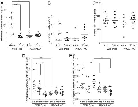
Fig. 1.
Comparison of testosterone and steroidogenesis levels during aging between wild-type and PACAP−/− animals. (A) Serum testosterone levels during aging in wild-type and PACAP−/− males. Between 4 (n = 8) and 15 (n = 8) months of age, serum testosterone decreased dramatically in wild-type animals, whereas levels remain low and constant in PACAP−/− mice (n = 9 for 4-month-old and n = 15 for 15-month-old animals) during the same period. (B and C) Comparison of serum LH (B) and serum FSH (C) levels during aging in wild-type and PACAP−/− males. Levels of LH and FSH hormones were assayed as described in Materials and Methods. For LH, 15 wild-type and 10 PACAP−/− males at 4 months of age and 6 wild-type and 9 PACAP−/− males at 15 months of age were used. For FSH, 16 wild-type and 10 PACAP−/− males at 4 months of age and 7 wild-type and 12 PACAP−/− males at 15 months of age were used. (D and E) Expression profile of StAR and 3β-HSD during aging. Testes of both wild-type (n = 5 for 4- and 8-month-old and n = 3 for 15-month-old) and PACAP−/− (n = 6 for 4-month-old and n = 5 for 8- and 15-month-old) animals were dissected and prepared as described in Materials and Methods. Level of expression of StAR (C) is significantly reduced in wild-type animals when aging but remained low and constant in KO animals. Similar experiments performed with 3β-HSD (D) revealed no significant difference in expression levels in wild-type animals during the aging process. In PACAP−/− mice, similar to that of StAR, the level of expression of 3β-HSD was low and constant over time. Data presented in the graph are the results of two independent real-time quantitative PCRs, performed with individual samples run in triplicate. ∗, P < 0.05; ∗∗, P < 0.01; ∗∗∗, P < 0.005.

