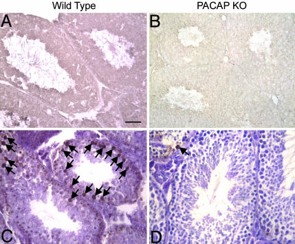
Fig. 3.
Peroxynitrite formation and testicular apoptosis in 15-month-old wild-type and PACAP−/− mice. (A and B) Peroxynitrite formation. Two testis sections of 15-month-old wild-type (n = 3) and 15-month-old PACAP−/− (n = 3) mice were stained with the anti-nitrotyrosine antibody to reveal the degree of peroxynitrite formation. Three different controls were included for each experiment: without the primary antibody, without the secondary antibody, and without primary and secondary antibodies, all showing a complete absence of staining (data not shown). Note the strong staining of peroxynitrites found in the old wild-type testis, in contrast to the moderate staining in the PACAP−/− testis. (Scale bar, 50 μm.) (C and D) Representative examples of apoptotic germ cells detected by the TUNEL method. In situ detection of cells with DNA strand breaks was assessed in three testis sections from 15-month-old wild-type (Left; n = 4) and PACAP−/− (Right; n = 5) mice, as described in Materials and Methods. For negative controls (data not shown), terminal deoxynucleotidyl transferase enzyme was substituted with PBS. Sections were counterstained with methyl green. Black arrows show the localization of apoptotic cells in the seminiferous tubules. (Scale bar, 50 μm.)

