Fig. 4.
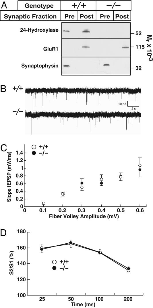
Synaptic transmission in WT and 24-hydroxylase KO mice. (A) The indicated hippocampal membrane fractions from WT (+/+) and KO (−/−) mice were probed for 24-hydroxylase, the GluR1 glutamate receptor subunit, and synaptophysin. (B) Spontaneous miniature synaptic currents in single CA1 pyramidal neurons in the presence of 1 μM tetrodotoxin. Representative traces from recordings made from 11 +/+ (○) and 14 −/− (•) neurons. (C) Input-output curves. Field potentials were determined in WT (○, n = 14) and KO slices (•, n = 13) over a stimulus intensity range of 5 to 40 μA. (D) Short term plasticity assessed by paired-pulse facilitation. Data from two different experiments with 43 +/+ (○) and 28 −/− (•) slices.

