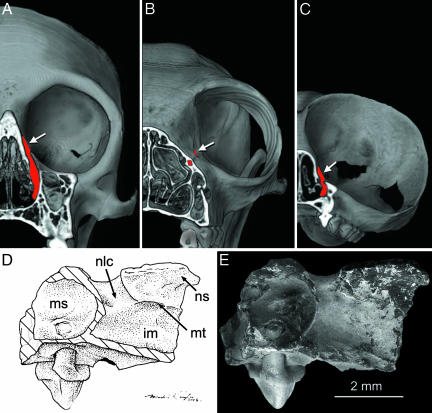
Fig. 3.
Internal anatomy of T. eocaenus. 3D computed tomography reconstructions of Cebus (A), Eulemur (B), and modern Tarsius sp. (C) compared with line art (D) and an SEM image (E) of T. eocaenus in medial view. The computed tomography reconstructions of modern skulls are cut in a coronal plane to show the course of the nasolacrimal ducts (filled in red). White arrows indicate their orbital opening. Note that in Eulemur the orbital opening is anterior to the orbit, and the duct travels horizontally, instead of vertically, so that the nasal opening is located much farther anterior than the plane of dissection. im, inferior meatus; ms, maxillary sinus; n/c, nasolacrimal duct.

