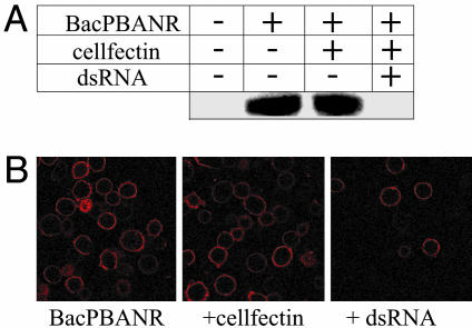
Fig. 2.
Silencing of recombinant PBANR. (A) Western blot analysis. BacPBANR-infected BmN cells were transfected at 3 h.p.i. with cellfectin ± 50 nM PBANR dsRNA overnight. At 48 h.p.i., cell lysates were immunoblotted and probed with an anti-His antibody. Uniformity of protein loading was confirmed by Coomassie stain. (B) Binding of a fluorescent PBAN analog. BmN cells treated as before were incubated with 50 nM rhodamine red-labeled PBAN for 1 h at 4°C. Cells were then fixed and imaged with a confocal fluorescence microscope.

