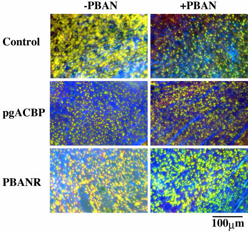
Fig. 4.
Accumulation and fluctuation of cytoplasmic lipid droplets. Images are pheromone-producing cells in the PG of 1-day-old decapitated females treated as pupae with DEPC-H2O (control), 5 μg of pgACBP dsRNA, or 10 μg of PBANR dsRNA. Images indicate ± PBAN treatment. Lipid droplets were stained with the fluorescent lipid marker, Nile red.

