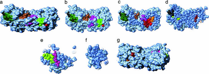
Fig. 5.
Putative GroEL SPBM (shown as contiguous-color regions) in the native conformation of Rubisco (light blue). (a and g) Front (back) view of the protein in a solvent-accessible surface area representation. (b–f) Cross sections, 10 Å apart, of the protein perpendicular to direction of observation in a. Molecular movies of the Rubisco surface and cross sections are available at www.biotheory.umd.edu/supplementary/motifs.html.

