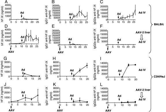
Fig. 1.
Plasma levels of hF.IX (A, D, G, and J), IgG1 (B, E, H, and K), and IgG2a (C, F, I, and L) anti-hF.IX in BALB/c (A–F) or C3H/HeJ (G–L) mice as a function of time. All mice (n = 4 per group) received i.v. injection of Ad-hF.IX at week 10 (marked “Ad”; 4 × 10¹⁰ particles per mouse). Mice were naïve at this time point (A–C and G–I) or had previously received hepatic AAV-hF.IX gene transfer (marked “AAV”) at day 0 (1 × 10¹¹ vg per mouse; D–F and J–L).

