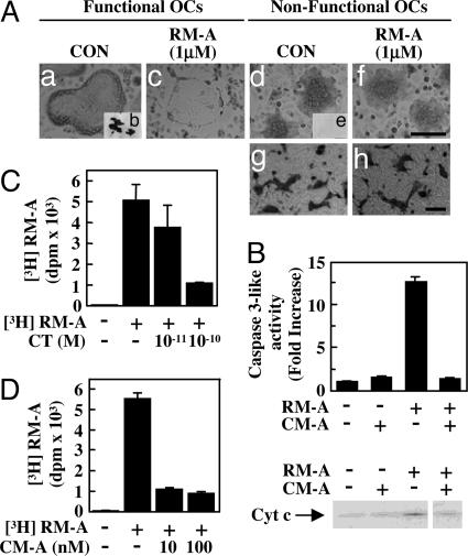
Fig. 2.
RM-A is incorporated into OCs. (A) TRAP (+) mononuclear osteoclasts prepared from cocultures (for 3 days) were cultured for 10 h on culture plates (Aa, Ac, Ad, and Af) and then treated for 8 h with or without RM-A (1 μM) in the presence or absence of RANKL (100 ng/ml) (Aa and Ac) or macrophage colony-stimulating factor (100 ng/ml) (Ad and Af). After culture, cells were fixed and stained for TRAP. TRAP (+) mononuclear osteoclasts were cultured on dentine slices (Ab and Ae) for 24 h. After culture, pits were stained with hematoxylin. (Scale bar: 10 μm.) Crude OCs prepared from cocultures were pretreated with CT (1 nM) and incubated with or without RM-A for 24 h. Cells were fixed and stained for TRAP (Ag and Ah). (Scale bar: 100 μm.) (B) Purified OCs were cultured for 1 h with or without CM-A (100 nM) in the presence of RANKL (100 ng/ml) and treated for 4 h with or without RM-A (1 μM). Cell lysates were used for measurement of caspase 3-like enzyme activity, and cytosolic extracts were used for Western blotting to detect cytosolic cytochrome c. Purified OCs were cultured with CT (C) or CM-A (D) for 1 h and then were treated with or without [3H]RM-A (5 μCi/ml) for 1.5 h. After treatment, cells were washed, and incorporation of [3H]RM-A into OCs was counted by using a liquid scintillation counter. Data are expressed as means ± SD for three cultures.

