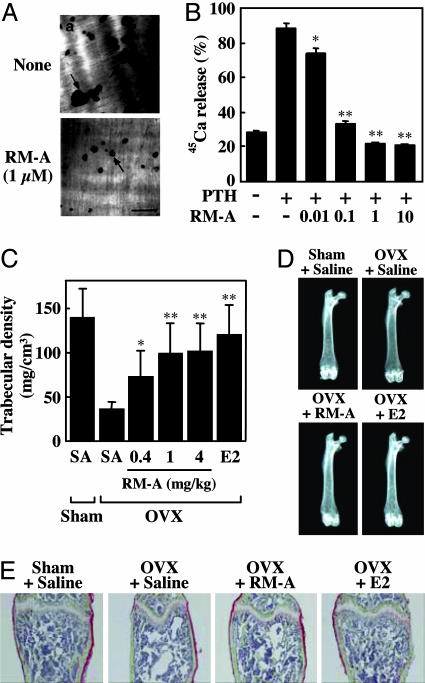
Fig. 4.
RM-A inhibits bone resorption in vitro and in vivo. (A) Crude OCs prepared from cocultures of bone marrow cells and osteoblasts were cultured on dentine slices for 24 h with (Ab) or without (Aa) RM-A (1 μM). At the end of the culture, resorption pits formed by OCs were stained with hematoxylin. Arrows indicate resorption pits. (Scale bar: 100 μm.) (B) 45Ca-labeled fetal rat limb bones were incubated with various concentrations of RM-A (0.01–10 μM) in the presence or absence of PTH (10 nM). Bone-resorbing activity was expressed as the percentage of incorporated 45Ca released into the medium. Data are expressed as means ± SD for four cultures. ∗, P < 0.05; ∗∗, P < 0.001 vs. PTH. RM-A (0.4, 1, and 4 mg/kg body weight twice daily), E2 (0.01 μg/day), or saline was administered s.c. to OVX mice starting 1 day after ovariectomy. After 4 weeks of treatment, mice were killed, and femora were removed for analysis of bone density and structure. (C) Trabecular bone density of distal femora was measured by peripheral quantitative computed tomography. Results were obtained from five mice per group and are expressed as means ± SD. ∗, P < 0.05; ∗∗, P < 0.01 vs. OVX plus saline. (D) Radiographic photographs of femora taken by soft x-ray. (E) Histological appearance of distal femora. Undecalcified bone sections were subjected to Villanueva Goldner staining (green, mineralized bone tissues). OVX + RM-A, OVX plus 4 mg/kg RM-A.

