
Figure 1.
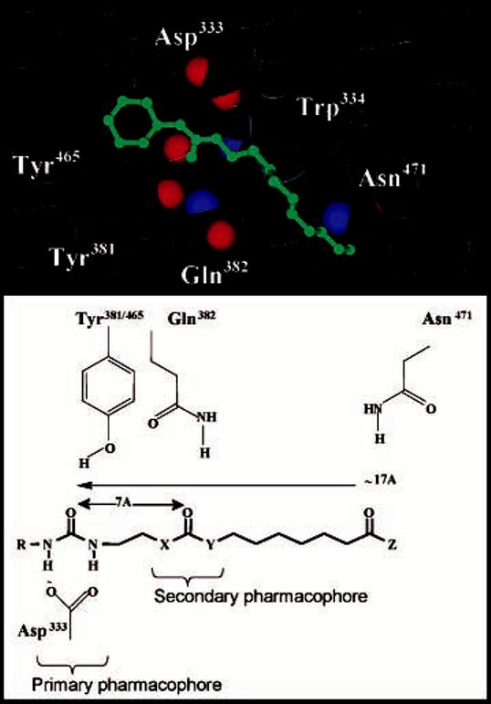
Diagram of the murine sEH active site and a sEH inhibitor: Distance of potential binding interaction from primary urea pharmacophore. The primary pharmacophore consistents of a polar carbonyl group, shown in this case as a urea with R = cyclohexyl, adamantyl, or 3-chlorophenyl. The secondary pharmacophore consistents of a polar group (ester, ketone, alcohol, or ether) located five/six atoms away from the carbonyl group of the primary pharmacophore.