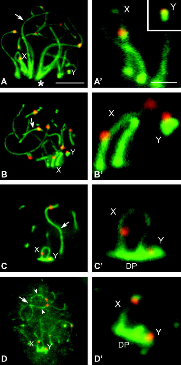
Figure 1.—

Immunolabeling of D. gliroides squashed spermatocytes with anti-SCP3 (green) and anticentromere (red) antibodies. Several optical planes of each nucleus were taken and subsequently superimposed. Only partial reconstructions of the nuclei are shown. (A) Zygotene. SCP3 appears on the autosomes as solid lines (arrow), whose ends are polarized toward a region of the nucleus (asterisk). Most autosomes are already synapsed. Sex chromosomes (X, Y) have well-defined AEs, but they appear located in opposite places in the bouquet area. (A′) Detail of the sex chromosomal AEs. (B and B′) Early pachytene. All autosomes are fully synapsed (arrow), but sex chromosomes still remain separated to each other in the nucleus, presenting thick and stiff AEs. (C and C′) Mid-late pachytene. Autosomes remain synapsed (arrow) and sex chromosomes are associated. The sex chromosomal AEs appear thinner than in previous stages and their labeling with anti-SCP3 is fainter. The DP labeled with anti-SCP3 is located on the tips of both sex chromosomal AEs. (D and D′) Diplotene. Autosomes desynapse and their LEs (arrowheads) appear separated all along the chromosome except at certain points (arrow). Sex chromosomes remain associated and the DP appears still organized. Bars: 5 μm for A–D; 1 μm for A′–D′.
