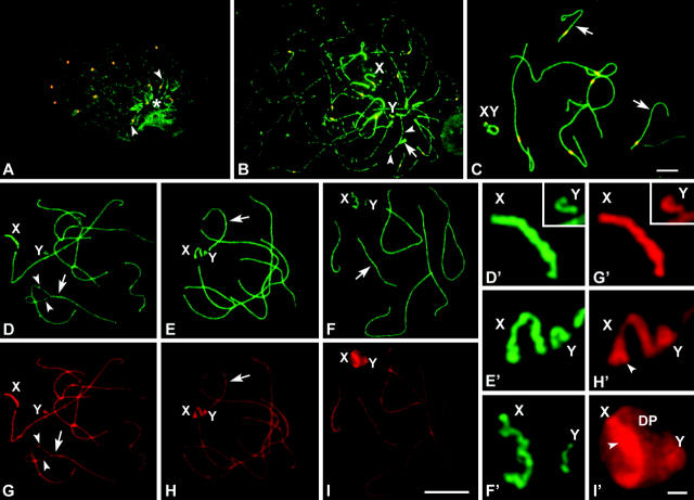
Figure 2.—
(A–C) Immunolabeling of spread spermatocytes of T. elegans with anti-STAG3 (green) and anticentromere antibodies (red). (A) Leptotene. STAG3 is present as small dots evenly distributed in the whole nucleus. These dots seem to condense to form linear structures (arrowheads) in a specific region (asterisk). All centromeres appear unpaired. (B). Zygotene. STAG3 appears forming irregular and interrupted linear structures. These lines are individualized in most of their length, but associate in pairs in some regions (arrow) and form fork-like structures. STAG3 appears in sex chromosomes (X, Y) as regular lines of similar thickness to the synapsed regions of autosomes. (C) Mid-late pachytene. The cohesin axes of autosomes are fully associated. Sex chromosomes appear associated, showing a regular labeling with STAG3, but there is no connection between the axes of the X and Y chromosomes. (D–I′) Immunolabeling of R. raphanurus spread spermatocytes with anti-STAG3 (green) and anti-SCP3 (red) antibodies. (D) Late zygotene. Autosomes are almost completely synapsed (arrow) and only some regions are still unsynapsed (arrowheads). Sex chromosomes (X, Y) lie apart from each other. (G) The same nucleus after SCP3 labeling. The localization of STAG3 and SCP3 is identical in both the synapsed and the unsynapsed regions of autosomes and sex chromosomes (enlarged in D′ and G′). (E and H) Midpachytene. Autosomes are fully synapsed and both proteins have a similar localization. However, the distribution of STAG3 and SCP3 is different on the sex chromosomes. STAG3 shows a linear pattern on both sex chromosomes, but this axial structure presents an irregular outline (E′). The outlines of the axes revealed by SCP3 are more regular and expand at the tips of both the X and Y chromosomes. (F and I) Late pachytene. STAG3 (F) and SCP3 (I) colocalize on the autosomes but present different patterns in sex chromosomes. While STAG3 reveals linear structures on the X and Y (F′), SCP3 not only reveals these axial structures but also appears in a plate-like structure in which sex chromosomal AEs are immersed (I′). This structure is the DP. Bars: 5 μm for A–C; 10 μm for D–I; 1 μm for D′–I′.

