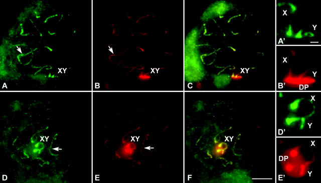
Figure 3.—
Immunolabeling of two pachytene squashed spermatocytes of T. elegans with anti-SMC3 antibodies (green) and anti-SCP3 (red). Several optical planes were taken of each nucleus and subsequently superimposed. (A) SMC3 is present along autosomes (arrow) as lines depicting the whole length of bivalents and is also present on the sex chromosomal axial elements (XY). (A′) Enlargement of the sex chromosomes. (B) SCP3 show the same pattern of distribution except for the sex chromosomes (XY), in which an intense labeling is detected in the region of association to the nuclear periphery, corresponding to the DP. (B′) Enlargement of the sex chromosomes. The DP is seen in lateral view. (C) Merge of SMC3 and SCP3 labeling. (D–F) A different spermatocyte, in which the DP is seen in polar view. Sex chromosomes are enlarged in D′ and E′. Bars: 5 μm for A–F; 1 μm for A′–E′.

