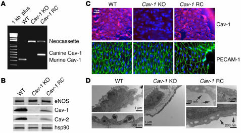
Figure 1. Characterization of carotid arteries from WT, Cav-1 KO, and Cav-1 RC mice.
(A) PCR genotyping from genomic DNA extracted from tails of WT (lower band, endogenous murine Cav-1), Cav-1 KO (upper band, neomycine cassette), and Cav-1 RC mice (middle band, canine Cav-1 transgene). 1 kb plus, DNA Mw ladder. (B) Protein levels of eNOS, Cav-1, and Cav-2 in 4 pooled carotid arteries. hsp90 was used as a loading control. (C) In situ whole-mount immunostaining performed in WT, Cav-1 KO, and Cav-1 RC carotid arteries showed expression of Cav-1 protein (red) and endothelial cell marker PECAM-1 protein (green). (D) Representative transmission electron micrographs performed in carotid arteries from WT, Cav-1 KO, and Cav-1 RC mice. Arrows indicate the presence of caveolae. C and D are representative of 4 experiments.

